The Coronary Artery Inflammation Controversy
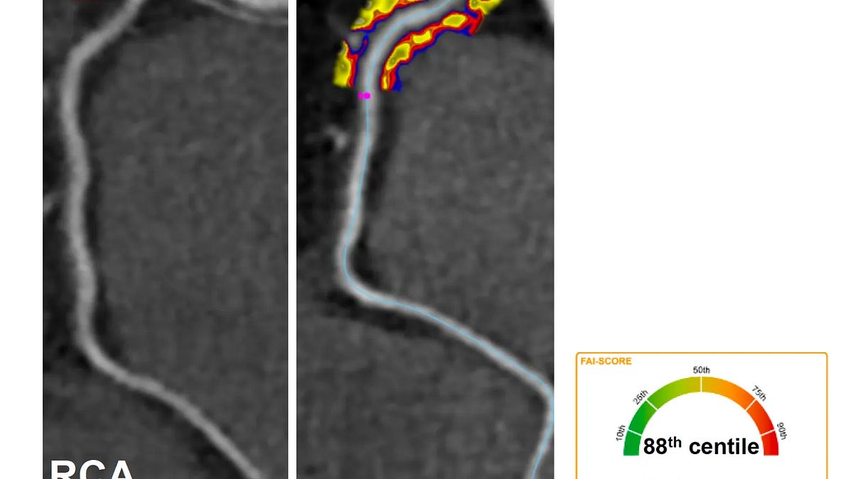
Why did a big clinical trial that tested an anti-inflammatory drug fail to reduce heart events? Why did another pivotal trial of lowering Lp(a) fail to reduce heart events? In this edition of Ground Truths I’ll offer some explanations and introduce some new and relevant information. In part, this is about relying on surrogate endpoints like increased blood levels of interleukin-6 or Lp(a), which have been generally regarded as an accurate reflection of heightened risk for coronary artery events. That may not be true. That these biomarkers are actually indicative of heightened arterial inflammation could be assessed using non-invasive AI quantitative assessment (FAI) of inflammation in the coronary arteries. That has now been done for high-sensitivity CRP (hs-CRP) and the results are informative. An example below is for a right coronary artery (RCA) that is not obstructed (red arrow) by atherosclerosis seen via a dye injection study (angiogram) but is actually highly inflamed (high FAI score right panel]. We’ll address each of the trials separately. First some general background on coronary atherosclerosis and inflammation. Some Background The coronary arteries have been center stage in recent weeks. Presented at the European Congress in Munich last weekend and published at NEJM, a prospective study of over 16,000 people aged 18-70 years found “silent atherosclerosis” (←no symptoms) in 57%, with many starting to show this at the young age of 18 to 29 years: 8.7% in men, 6.7% in women. Prior studies suggest the prevalence in young adults is considerably higher (~4-5 fold using intravascular ultrasound). In JAMA last week, an assessment of calcium scores of the coronary arteries by CT scans questioned their overall utility compared with clinical criteria, recommending selective use. Both new studies underscore the high incidence of coronary atherosclerosis, cholesterol-laden plaque, that is not accompanied by symptoms. An important differentiation: coronary atherosclerosis per se is extremely common, but plaque rupture leading to heart attacks (MI, myocardial infarction) or unstable angina is relatively infrequent (about a 50:1 ratio, US data, Figure below, citation). Why? We have known for decades, from the pivotal work of Russell Ross and others who did painstaking histopathology studies to trace the origin and evolution of coronary atherosclerosis, that inflammation plays a central role. From the panels top to bottom below, migration and adhesion of white blood cells are key in the early stage, T-cell activation in the next, then macrophage accumulation, culminating in plaque rupture in the bottom panel. The latter event is what leads to heart attacks. It doesn’t require a marked accumulation of plaque (known as “critical stenosis” or colloquially as a “blockage”) but instead can occur in an inflamed artery without much atherosclerotic burden. But these studies were based on pathology of arteries which are only accessible post-mortem or at the time of surgery. So we have not had a direct handle on the process of coronary artery inflammation. High-risk plaque for rupture isn’t the same thing, but that can be assessed by placing a catheter inside the artery using intra-coronary imaging tools (intravascular ultrasound, IVUS or optical coherence tomography, OCT). Without a non-invasive way to determine coronary artery inflammation, we’ve resorted to the use of blood biomarkers, such as high-sensitivity C-reactive protein (hs-CRP) and interleukin-6 (IL-6). As I’ve reviewed in prior Ground Truths, there were 5 randomized trials with colchicine in people with heart disease and increased hs-CRP which had an aggregate 25% reduction of heart attack, 46% reduction of stroke. There was also a large trial of an interleukin-1β antibody (vs placebo) in over 10,000 participants with heart disease and elevated hs-CRP. The 2 higher drug doses of the antibody led to about 15% reduction of the primary endpoint of heart attack, stroke or cardiovascular death. But these biomarkers are notoriously non-specific, with any type of inflammation in the body leading to their elevation. Think joint inflammation or a common cold. Further, the magnitude of efficacy for these two interventions (colchicine and the antibody) was considered low with a trade-off of significant side effects, like increased serious infections with the interleukin-1β antibody. But, in 2025, these findings along with new insights about vulnerable plaque and vulnerable patients, as summarized in The Big Shift to Inflammation, prompted the American College of Cardiology (pull quote below) and a Lancet Commission to publish the new objective of targeting coronary artery inflammation and early detection of atheroma. “The time is also ripe for the development of strategies to promote increased physician awareness of the crucial role of inflammation in CVD and accelerate the adoption of evidence-based, guideline-directed anti-inflammatory therapy through dissemination and implementation research.” Enter ZEUS While not yet published, Novo Nordisk, the sponsor of the >6,300 participant trial of ziltivekimab, an IL-6 antibody trial vs placebo, announced the results at the end of July by press release. The inclusion criteria were people with atherosclerotic vascular disease and chronic kidney disease, and hs-CRP level of 2mg /L or higher. The drug, 15 mg per month, markedly reduced the levels of IL-6 and hs-CRP but there was no reduction of cardiovascular death, nonfatal MI or nonfatal stroke. This led to coverage in the media, such as the New York Times (headline below) to declare “its shocking failure” and question “whether inflammation really does cause heart disease.” Incidentally, at the Munich ESC over the weekend, another IL-6 monoclonal antibody (pacibekitug) trial was presented that showed a dose-dependent reduction of IL-6 levels but was much smaller and did not report clinical outcomes. These are just 2 of a large group of IL-6 antibodies that include clazakizumab, olokizumab, siltuximab, sirukumab), IL-6 receptor blockers, including tocilizumab, sarilumab, satralizumab, vobarilizumab, and novel blockers such as an IL-6 trap or combination blockers with one countering IL-6. So there’s a lot on the line for the biopharma world invested in this specific target of anti-inflammatory drugs. Why Zeus Failed? There are a number of possibilities but this one— there’s something wrong with the “inflammation hypothesis” —is way off the mark. Marios Georgakis wrote an outstanding essay on the topic offering a differential diagnosis for the failure that included problems with IL-6 as the target, that the IL-6 receptor is a better target, or the wrong population was studied— specifically the inclusion of people with chronic kidney disease who are well known to have an elevated hs-CRP, IL-6 and other biomarkers of heightened inflammation. I will be discussing this very important topic and the Lp(a) trial results with Prof Georgakis on a live video on September 9 at 9 AM PDT so please join us. That gets me to why I believe Zeus failed to show clinical benefit, in part related to the chronic kidney disease inclusion. The issue is having elevated hs-CRP and IL-6, very non-specific markers, WITHOUT coronary artery inflammation. None of the patients in Zeus had their coronary arteries assessed for inflammation, which is now possible and a big miss for enrolling participants who could potentially derive benefit. Further dilution of any potential benefit was achieved by enrolling people with chronic kidney disease (without prior heart disease) who are well known to have abnormal inflammation biomarkers unrelated to coronary artery disease. Non-Invasive Assessment of Coronary Artery Inflammation As I wrote in The Big Shift in Cardiology to Atheroma and Inflammation we can do this with AI of the epicardial (tissue surrounding the artery) fat to quantify the level of inflammation for each of the 3 coronary arteries. This was validated by the University of Oxford team of researchers in 40,000 consecutive patient CT angiograms with long term clinical follow-up and with histologic correlation of post-mortem arteries. Back to the graphic I used at the top of the post, the acronym term FAI is for fat attenuation index, the coronary AI score, and below it what it looks like (red and blue indicate increased inflammation) for a right coronary artery with no significant obstruction (but is highly inflamed). Inflammation proved to be exceedingly important, independent of obstruction (high grade stenosis) as seen below. We hardly ever see a hazard ratio of 13-fold for any marker, as was seen for one inflamed coronary artery predicting cardiac death during 10-year follow-up. No less ~30-fold risk for all three arteries inflamed! They spun out a company from University of Oxford called Caristo Diagnostics which received FDA authorization on July 29th, 2026—’”the first and only technology authorized to quantify coronary inflammation from routine coronary CT angiography.” This is distinct from the other companies that assess plaque or physiology but not inflammation (see link, Table below) To note, I have no financial relationship with this company. I have been very interested in this emerging capability and I’ve been writing about it (the GT links above) for the past 2 years. Now it’s even more relevant. The Oxford team has rich data to answer the question does hs-CRP level correlate with coronary artery inflammation? The answer is no. As presented at the ESC Madrid in 2025, the correlation of FAI score and hs-CRP is poor, r = 0.2. We have no knowledge as to whether the participants in ZEUS had coronary inflammation even though there was evidence of systemic inflammation by increased circulating blood levels of IL-6 and hs-CRP. Furthermore, although the IL-6 blocker led to pronounced reduction of IL-6 levels, as expected, that doesn’t establish it was related to any decreased coronary inflammation. This essential point was reinforced at the Munich ESC meeting from a state-of-the-art ongoing trial of a monoclonal antibody vs oxidized LDL cholesterol (orticumab) that assessed coronary artery inflammation by CT angiography before enrollment and drug treatment. In 512 screened participants, the hs-CRP was normal (0.8 mg/L) but 63% had an FAI score at or above 50% in one or more coronary arteries. Another way to say all of this, if you want to test whether a drug reduces coronary inflammation, you need to know whether inflammation is present in the coronary arteries! Using hs-CRP or IL-6 is not an adequate surrogate marker. What is especially noteworthy about FAI is that until recently it required a coronary CT-angio, that is an intravenous injection of dye and more radiation and expense to acquire the images and apply the AI scoring. Last month the Oxford investigators, from >19,000 patients with 6.8 years of follow-up, showed the FAI from a non-contrast chest CT worked just as well. The contrast and non-contrast FAI results were tightly correlated (as seen below), as were the risk of subsequent major cardiovascular events. This makes the use of a low cost chest CT a very practical way to assess and quantify coronary artery inflammation. Enter HORIZON We’ve know that at least 20% of people have an elevated Lp(a), a genetic trait not influenced by lifestyle factors to any significant degree. High levels of Lp(a) are incontrovertibly correlated with increased risk of coronary artery disease, heart attacks, stroke, and peripheral vascular disease. Just this week a new study using genetic analyses with Mendelian randomization showed that combined lowering of LDL and Lp(a), in people with genomic risk of these elevated lipoproteins, was linked to improved outcomes. How does the increased Lp(a) accelerate atherosclerosis? It’s an apoB- containing particle that takes up residence in the arterial wall (Schematic Figure above stages 1 and 2) and has oxidized phospholipid (OxPL) cargo that is highly pro-inflammatory (Figure below), far greater than LDL cholesterol. Its structure is akin to plasminogen and that may, in part, explain why it also increases propensity for clotting. An Lp(a)blood measurement provides no information about OxPL. For decades we have known about the risk but were without any known treatment. All we could do was to be aggressive with LDL cholesterol lowering and consider anti-platelet therapy with low dose aspirin. Then, finally, came various drugs that could markedly and safely lower Lp(a) which was confirmed in Phase 2 trials. But would they lower outcomes? The first outcome trial for Lp(a) reduction was with the antisense oligonucleotide pelacarsen and was also presented only by press release. This was a classic randomized, double-blind multinational trial with >8,300 participants and over 900 sites that initiated enrollment in 2019 and has been the frontrunner for all the Lp(a) trials. Parenthetically, I would add it was interesting the press release was done on a Friday evening instead of during the recent ESC Munich conference less than a week ago, but that may simply be coincidence of when the data were ready to make an announcement. We’re missing many details that are not available about the demographics of the participants , how well their LDL cholesterol was treated, no less the results for subgroups (like those with the most extensive Lp(a) reduction or the highest clinical risk) and so much else. So why did it fail? There are many possible explanations such as a lower risk participant population by clinical criteria or were on maximal medical therapy. The one that I find particularly likely is that Lp(a) as a marker is non-specific. Unlike IL-6 or hs-CRP which can be high in the blood but not denoting inflammation in arteries, Lp(a) is known to be most pro-inflammatory when there are high levels of oxidized phospholipid (OxPL, its cargo), which are not measured clinically (or in the clinical trials). So some people with a high Lp(a) do not have arterial inflammation while others do. And it’s not binary, but a continuum. In parallel, we learned this about elevating HDL in clinical trials with niacin and other drugs. Low blood levels of HDL correlated well with increased cardiovasular risk but raising it did not affect clinical outcomes. Then we became aware that levels of HDL are more complicated, different functionally, and that even very high levels of HDL are linked with increased risk. It’s about the default of using a lipid metric that is not broken down with respect to its actual pathogenicity (fancy word for causing disease). Like OxPL cargo for Lp(a). There are other possible explanations. The drug used in Horizon was an antisense oligonucleotide (ASO), not as potent as some of the others (Table) and unlike siRNA (small interfering RNA drugs); it’s half-life is shorter and has to be given more frequently. The inclusion criteria for Lp(a) elevation was somewhat reduced in HORIZON, but note the difference in units: nmol/L — nanomoles per liter particle concentration (in all the other programs and the current preferred lab readout) vs the traditional metric mg/dL — milligrams per deciliter for mass concentration, no used by much labs these days. The HORIZON trial Lp(a) mg/dL entry would be equivalent to ~150 nmol/L. So we’ll have to see whether the other programs provide different results for outcomes. That will help dissect these uncertainties with respect to potency, entry criteria, and type/dosing of drug. We’ll be left in suspension for some time since the estimated completion of the next trial, olparisan, is not until 2028; the others between 2029-2031. That shows how far ahead Novartis was as the pacesetter for pelcarsen. The Bottom Line We can’t count on surrogate blood markers to guide the results of cardiovascular clinical outcome trials. We’ve seen this multiple times over past decades with interventions that raise HDL or lower triglycerides. It’s not so simple a translation from a blood test to reducing heart attacks and strokes as worked, in general, for lowering LDL cholesterol. The “inflammation hypothesis” is not a hypothesis. It has been rigorously proven by multiple modalities, pathology, and clinical studies to be a critical component of atherosclerotic coronary artery disease and its propensity to result in heart attacks and clinical events. What hasn’t been adequately established is a safe potent intervention to reduce the arterial inflammation. That is not going to be established by relying on circulating blood biomarkers since they are clearly dissociated from what is happening in the coronary arteries. While there are abundant blood tests for lipids [LDL, ApoB, HDL, Lp(a)] we don’t have any surrogate blood markers for coronary inflammation. The Lp(a) metric is especially complicated because it doesn’t get to the actual pro-inflammatory, pathogenic potential. I should add that there are many patients who have completely normal lipids but have coronary inflammation and develop heart attacks. Our undue emphasis on abnormal lipids as the only culprit misses this subpopulation of people at risk. Fortunately, we do have a non-invasive method to identify and quantify coronary inflammation with AI. We know that arteries with a calcium (CAC) score of zero can be highly inflamed, and that high CAC scores are not necessarily high-risk for cardiovascular events. Over the years, the cumulative number of patients referred to me for a high CAC score, without symptoms, and demonstrated by CT or coronary angiography to have no significant coronary artery obstructive disease is in the hundreds. While I have been against the use of calcium score yet very supportive of coronary CT angiograpy when indicated for select patients, this is likely to change. The CT that could only provide data on calcium, which does not reflect obstructive disease in many patients, will soon be AI interpretable for presence and severity of coronary inflammation. Trials of future anti-inflammation drug candidates will be able to get a low cost chest CT and enroll participants who actually have evidence of coronary artery inflammation. That has exciting potential to help catalyze our hunt for safe and potent drugs. Until the right trials are done, with enrollment of patients with heart disease, not kidney disease, with bona fide coronary inflammation, it would be misguided to refute such a compelling body of evidence. Finally, what should you do if you have a high Lp(a), such as >175 nmol/L? Use all the factors we know to lower risk, like lifestyle factors of diet, exercise, and medication lowering of LDL, apoB, along with blood pressure control. Are these measures working to mitigate risk in a person? Since there’s a long wait time to know about the other Lp(a) drugs in the pipeline, perhaps knowing about coronary artery inflammation will turn out to be helpful when plain CT FAI oo OxPl blood tests becomes widely available (assuming very low cost). NB. This post was written by me without any contribution from AI. I have no conflict of interest related to this post and specifically, as mentioned above, no relationship with Caristo Diagnostics. Ground Truths has 215,000 subscribers from every US state and 214 countries. There are over 300,000 followers of Ground Truths so more than 90,000 folks who can easily convert to be free subscribers. Your subscription to these free essays and podcasts makes my work in putting them together worthwhile. If you’re not a subscriber, please join! If you found this interesting PLEASE share it! The proceeds from all voluntary paid subscriptions go to support our summer internship program. It enabled us to accept and support a record number of 62 summer interns that joined us in 2026! These are high school, college and medical students selected from thousands of applicants. We couldn’t do this expanded program without the funds coming in through Ground Truths. Thank you!
~14 min read · 3156 words